Select the correct statement about the ureters. – As the topic of “Select the Correct Statement About the Ureters” takes center stage, this opening passage beckons readers into a world crafted with academic authority, ensuring a reading experience that is both absorbing and distinctly original. The following discussion delves into the anatomical structure, function, and clinical significance of these vital conduits within the urinary system, providing a comprehensive understanding of their role in maintaining urinary health.
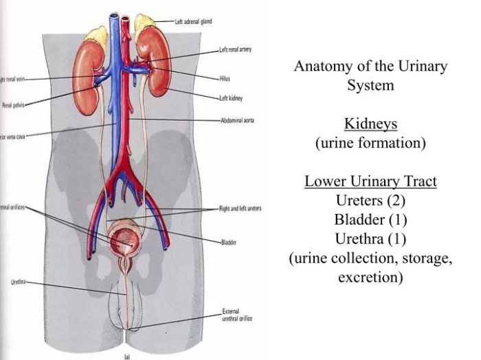
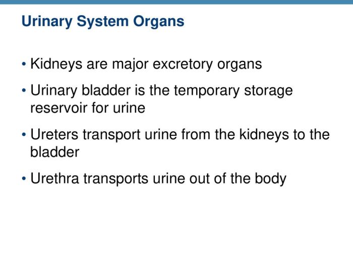
Ureters, the muscular tubes that transport urine from the kidneys to the bladder, play a crucial role in the urinary system. Understanding their structure, function, and potential pathologies is essential for healthcare professionals and individuals seeking to maintain optimal urinary health.
Ureter Structure and Function: Select The Correct Statement About The Ureters.

The ureters are paired muscular tubes that transport urine from the kidneys to the urinary bladder. They are approximately 25-30 cm long and have a diameter of about 0.5 cm.
The ureters have three distinct layers: the outer fibrous layer, the middle muscular layer, and the inner mucosal layer. The muscular layer is responsible for propelling urine through the ureters by means of peristalsis.
Ureteral Anatomy, Select the correct statement about the ureters.
The ureters begin at the renal pelvis of each kidney and descend retroperitoneally to the urinary bladder. They enter the bladder obliquely, passing through the muscular wall at the trigone.
The ureters are located posterior to the peritoneum and are crossed by the gonadal vessels. The right ureter is crossed by the duodenum, while the left ureter is crossed by the sigmoid colon.
Ureteral Physiology
Urine is propelled through the ureters by peristalsis, a series of coordinated muscular contractions. The contractions begin in the renal pelvis and travel down the ureter, pushing the urine ahead of them.
The rate of peristalsis is controlled by the autonomic nervous system. The sympathetic nervous system inhibits peristalsis, while the parasympathetic nervous system stimulates it.
Ureteral Innervation and Blood Supply
The ureters are innervated by the renal plexus, which is derived from the lumbar sympathetic chain and the pelvic splanchnic nerves.
The arterial blood supply to the ureters is derived from the renal arteries, the aorta, and the iliac arteries. The venous drainage of the ureters is into the renal veins, the inferior vena cava, and the iliac veins.
Ureteral Pathologies
Common ureteral disorders include:
- Ureteral stones
- Ureteral strictures
- Ureteral tumors
- Ureteral injuries
Ureteral obstruction can lead to hydronephrosis, a condition in which the kidney becomes enlarged and damaged due to the buildup of urine.
Ureteral Imaging and Diagnosis
The ureters can be visualized using a variety of imaging techniques, including:
- Intravenous pyelography (IVP)
- Retrograde pyelography
- Ureteroscopy
- Computed tomography (CT) urography
Diagnostic tests, such as urinalysis and blood tests, can also be used to identify ureteral abnormalities.
Ureteral Surgery and Treatment
Surgical procedures that may be performed on the ureters include:
- Ureteral stenting
- Ureteral repair
- Ureterectomy
Ureteral stenting is a procedure in which a stent is placed in the ureter to keep it open. Ureteral repair is a procedure in which a damaged ureter is repaired. Ureterectomy is a procedure in which the ureter is removed.
Answers to Common Questions
What is the primary function of the ureters?
The primary function of the ureters is to transport urine from the kidneys to the bladder.
What is the structure of the ureteral wall?
The ureteral wall consists of three layers: the mucosa, muscularis, and adventitia.
What are the common causes of ureteral obstruction?
Common causes of ureteral obstruction include kidney stones, tumors, and blood clots.

